
Pro l kandidiasis pada pasien yang berkunjung ke klinik. Ciri khas lesi berbentuk seperti jala meny ilang dikenal sebagai wickham striae bersifat kronis dapat terjadi pada kulit mukosa atau kulit dan mukosa.

Gambar 5 Perbaikan Kondisi Intra Oral Pasien Kedua Setelah Satu Minggu Download Scientific Diagram
It can be isolated from alm ost 50.
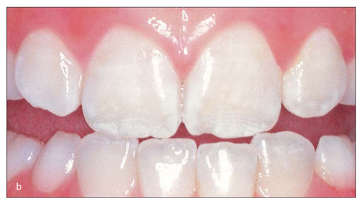
. Oral white lesions can be caused by a thickened keratotic layer or an accumulation of non-keratotic material. LESI PUTIH HEREDITARY WHITE. Extended use of microwave ovens has caused an increased prevalence of thermal burns since they heat up food unevenly in a way that the inner portion of it remains cold while the other part becomes hot 4514.
In many cases the lesion can be classified as recurrent herpes labialis but many other causes can induce a vesiculobullous lesion of the oral mucosa and perioral skin as well. Asymptomatic most of the times except when there is erosion of the lesion. Thermal burns of the oral cavity generally result from ingestion of hot foods or beverages like hot pizza or coffee.
Oral cavity such as intubation during general anaesthesia. Linea Alba Linea alba is a common finding which is. Buccal mucosa is the most common location tongue gingiva and palate are the less common locations.
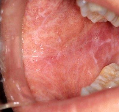
White Striae are seen. Chemical Burn Perubahan lesi putih nonkeratotik sering diakibatkan oleh injuri kimia ketika agen kimia berkontak dalam waktu yang cukup lama. Diagnosis of oral white lesions might be quite challenging.
Similarly chemical or physical injury could result from undue or careless handling of chemicals and dental instruments during dental treatment procedures. They may spread as diffusely or remain consolidated in one particular site. Lesi Putih biasanya dapat berupa plaque atau papula.
General search engines and specialized databases including PubMed PubMed. Kondisi Herediter Leukoedema White sponge nevus Hereditary benigna intra-epithelial dyskeratosis HBID dan Keratosis. Clinical Features of White Lesions of the Oral cavity.
Lichen Planus Clinical Features. No specialised classification of lesion. This article gives an overview of the various vesiculous.
Textbook of Oral Medicine Oral Diagnosis and Oral Radiology. Bilateral seen on both sides of the oral cavity. Agen tersebut dapat berupa aspirin silver nitrate formocresol sodium hypochlorite paraformaldehyde dental cavity varnishes acidetching materials dan hydrogen peroxide.
This article presents the most recent information related to the management of several types of white lesions of the oral cavity. Lokasi paling sering ditemukan lesi plak pseudomembran putih dan terdapat di daerah dorsal lidah. Lesi putih oral cavity.
Lesi Putih liken planus yang disertai lepuh mulut. Are You Missing the Diagnosis a Critical Images slideshow to help identify the causes of abnormalities of the oral cavity. Pigmentations in the oral cavity vary from color to color and also in shape and size.
If so a superficial non-keratotic layer such as pseudomembranes most commonly caused by. Zc½1 åtë bhaäl Ãß ºd 47U7dM lTé. See Clues in the Oral Cavity.
Gambaran Klinis Epitel displasia dan karsinoma sel skuamosa pada stadium awal memiliki gambaran klinis berupa bercak putih fokal sekitar mukosa oral yang secara superfisial terlihat serupa dengan lesi hiperkeratosis. Apa itu lepuh mulut. This paper reviews some common causes of traumatic injuries their diagnosis and management Table 1.
Munculnya lesi berwarna putih dapat disebabkan oleh karena perubahan. Of the oral cavity and oropharynx w fibroelastic change and inflammation of the mucosa progressive inability to open the mouth swallow or speak Very resistant to treatment be a premalignant condition oral cancer developed in 76 of patients malignant transformation rate was 4 to 13. Classification Oral pigmentation has been associated with variety of lesions and conditions.
Oral pigmentation is a relatively common condition that may involve any part. Lesi putih oral cavity What Is Oral Lichen Planus News Dentagama What Is Oral Lichen Planus News Dentagama Crown Pfm Close Up Of Molluscum Contagiosum Also Called Water Wart Stock Photo Image Of Fluconazole Health 132122748 Gambar 2 Gambaran Klinis Intra Oral Setelah Satu Minggu Pengobatan Download Scientific Diagram Gambar 5. In general practice the dentist can be confronted with a vesiculobullous lesion of the oral mucosa.
They can occur either under normal conditions. Wound ulcer abscess or tumour. Lesi putih berupa Oral lichen planus.
Pemeriksaan klinis secara detil akan menunjukkan tekstur permukaan dari lesi displasia lebih kasar atau berkerut setelah dilakukan pengeringan pada. Lesi Putih presentasi - Free download as Powerpoint Presentation ppt PDF File pdf Text File txt or view presentation slides online. An area of abnormal tissues.
A region in an organ or tissue which has suffered damage through injury or disease. Lesi Putih dapat dibedakan menjadi. Regezi Sciubba 1993 1.
Accordingly when a clinician confronts a white area on the oral mucosa the first issue to be elucidated is whether it can be scraped off by means of a piece of gauze or not. Naming by their pattern size location or causes. Oral candidiasis is the most common fungal infection of the oral cavity mostly caused by Candida Albicans as one of the normal microflora organisms 41 6.
Lepuh mulut merupakan suatu poket kecil yang berisi cecair di permukaan superfisial mukosa mulut. Lepuh jarang dijumpai di dalam mulut kerana ia senang pecah dan menjadi ulser mulut kerana persekitaran yang lembap di dalam mulut juga daripada trauma semasa mengunyah makanan. This review article aimed to introduce a decision tree for oral white lesions according to their clinical features.
Lesi putih yang terbentuk. The most commonly affected areas are palatal mucosa. Lesi Putih pada mukosa rongga mulut Area keputihan - mukosa oral - khas ok pencahayaan.
Candidiasis has a variable presentation but can consist of slightly elevated. Any damage or abnormal change in the tissue of an organism. LESI PUTIH 50 dari lesi jaringan lunak yang ada di rongga mulut seluruhnya berwarna putih atau memiliki beberapa komponen yang berwarna putih.

Epstein Pearls In Infants What Causes And How To Treat

Lesi Mulut Tanda Dan Gejala Penyebab Cara Mengobati Cara Mencegah

Pin On Instagram Doktergigigaul

Pin On Instagram Doktergigigaul
3 Partial Tooth Discolorations Pocket Dentistry
What Is Oral Lichen Planus News Dentagama
2

Diagnosis Recurrent Aphthous Stomatitis Alomedika

Close Up Of Molluscum Contagiosum Also Called Water Wart Stock Photo Image Of Fluconazole Health 132122748
2
Lesi Dan Gejala Pada Rongga Mulut Penderita Covid Unair News

Pin On Instagram Doktergigigaul

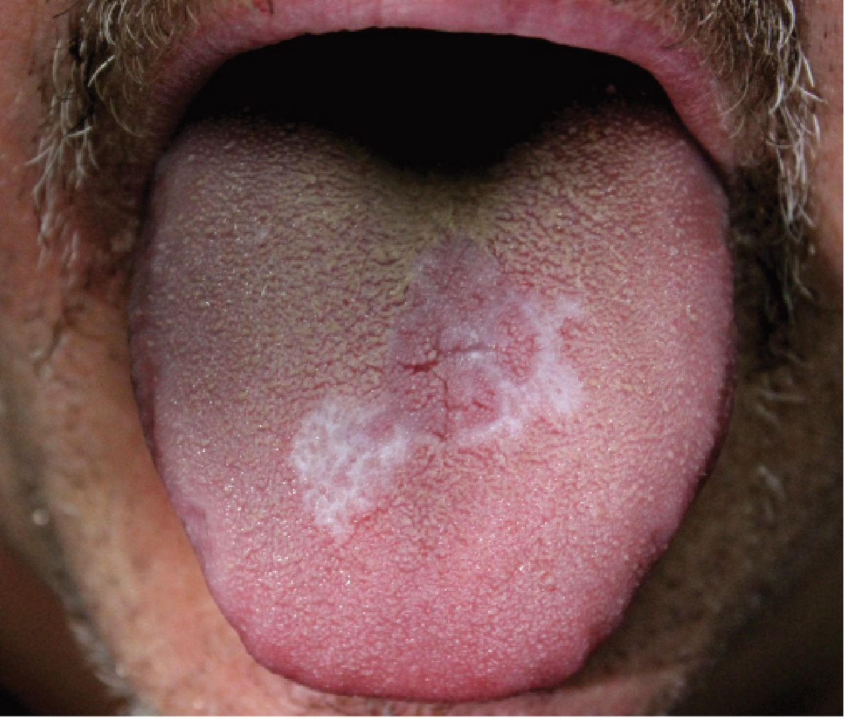
What On Earth Is Your Tongue Trying To Tell You Ever Wonder Why The Doctor Asks You To Open Your Mouth A Geographic Tongue Glossitis Burning Tongue Syndrome

Ulcer On The Left Buccal Mucosa Ovoid In Shape Download Scientific Diagram
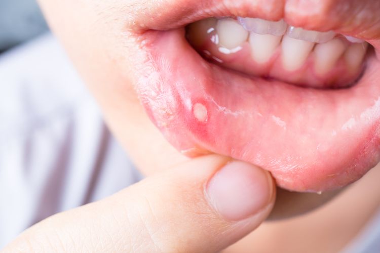
5 Cara Membedakan Sariawan Dan Kanker Mulut Halaman All Kompas Com
![]()
Extensive Ulceration And Characteristic Crust Formation Arrow On The Download Scientific Diagram
Oral Hairy Leukoplakia Sebagai Prediksi Lesi Rongga Mulut Untuk Penyakit Hiv Unair News
What Is Oral Lichen Planus News Dentagama

Ulcer With Yellow Crusted Surface On The Left Posterolateral Border Of Download Scientific Diagram


